Other Knee Ligament Tears: MCL & LCL
Medial collateral ligament (MCL)
MCL injury is the most frequent knee ligament injury. Fortunately, it heals well without the need for surgery
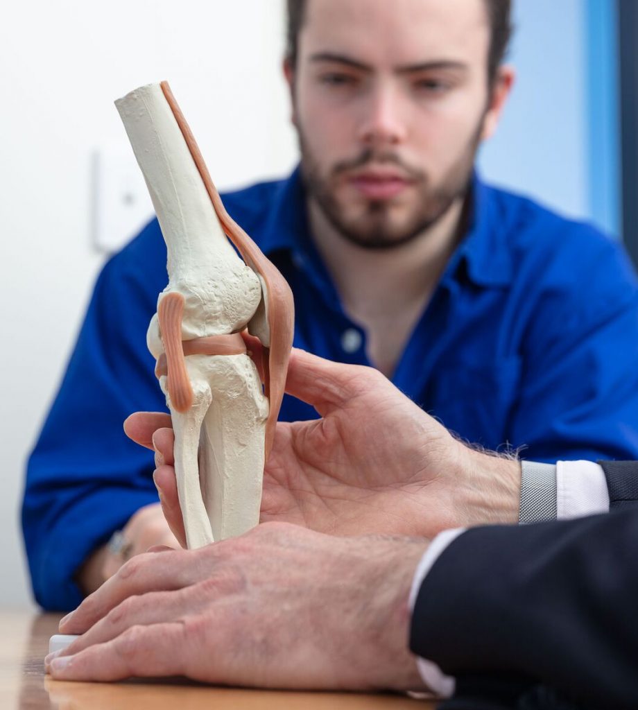
The MCL is a broad and long ligament that arises from the medial (inner) epicondyle at the end of the femur and runs to the upper part of the tibia. The function of the MCL is to prevent sideways movement of the knee. A direct blow to the outside of the knee or an indirect force will tear the MCL.
Ligament sprains are classified according to the severity. A Grade 1 sprain is a mild injury with no instability. Grade 2 sprains are partial tears, and the ligament feels loose. Grade 3 sprains are a complete tear.
Treatment
Nonoperative management will heal all isolated MCL injuries. The initial first aid is to rest the knee, apply compression and ice to reduce swelling. Bearing weight is allowed, but a higher grade injury may require the use of crutches for a period. Early movement as pain permits is vital to prevent knee stiffness. Higher grade injuries require a brace to protect the ligament while healing. When the tear heals a strengthening program can commence. When pain-free and the ligament is demonstrably stable, the person may return to sport.

It is unusual for MCL sprains to cause any chronic (after three months) problems. Occasionally people can have pain because of abnormal calcification where the damage had been. Also, uncommonly people can have instability followed a complete tear. They, therefore, may require an MCL reconstruction.
Dr Brown provides consultation and treatment for MCL, PCL and LCL
Call us on (03) 5223 3151 Book an appointment today.
Lateral collateral ligament (LCL)
Often the LCL is torn in association with other ligament injuries, particularly the ACL or the PCL
Treatment
Nonoperative management will heal Grade 1 and 2 sprains, as for MCL injuries. Complete tears of the LCL are usually associated with other local ligament damage and don’t heal. Therefore these injuries need early assessment with a thorough knee ligament examination and an MRI scan. Typically repair or reconstruction within two weeks from injury is required.


Posterior cruciate ligament (PCL)
The PCL is a robust ligament that runs from the tibia to the femur.
The PCL prevents the tibia from moving excessively backwards. In the sporting context, the PCL tears with a hyperextension injury such as landing from a jump. Contact by another player may forcibly drive the tibia backwards. In Australian Rules football, it is common in ruckman from the clash of knees during a ball up. The other classic injury pattern is the “dashboard injury” that occurs in a motor vehicle accident. Here the tibia hits the dashboard, and the strong impact forces it backwards.
Treatment
Isolated PCL tears are usually treated non-operatively even in elite athletes. Usual first aid measures are used, such as described as above. If diagnosed early, a specific PCL brace prevents the backwards sag of the tibia, facilitating healing. Early movement in the splint is essential. A strengthening program commences once healing is underway. Strengthening of the quadriceps or “thigh” muscle is critical as it has an action of pulling the tibia bone forwards, therefore, counterbalancing the posterior sag.
PCL repair or reconstruction is sometimes required. If the PCL and another ligament tears, early surgery is indicated. If the PCL tears off the bone, then it can be repaired. If the PCL suffers a mid-substance rupture, then it will need to be reconstructed. Some PCL tears result in ongoing instability or pain. These chronic tears may require a reconstruction as well.

